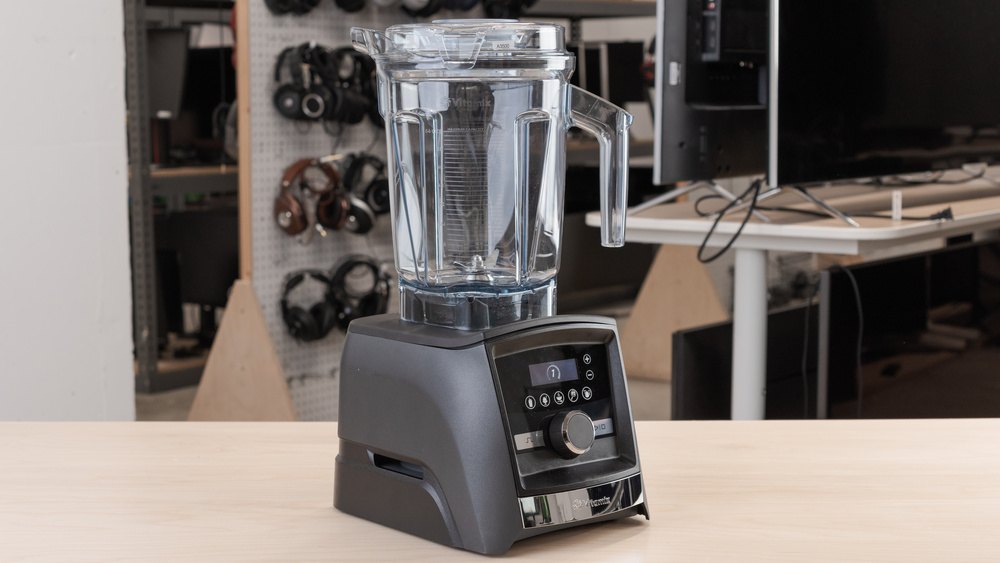
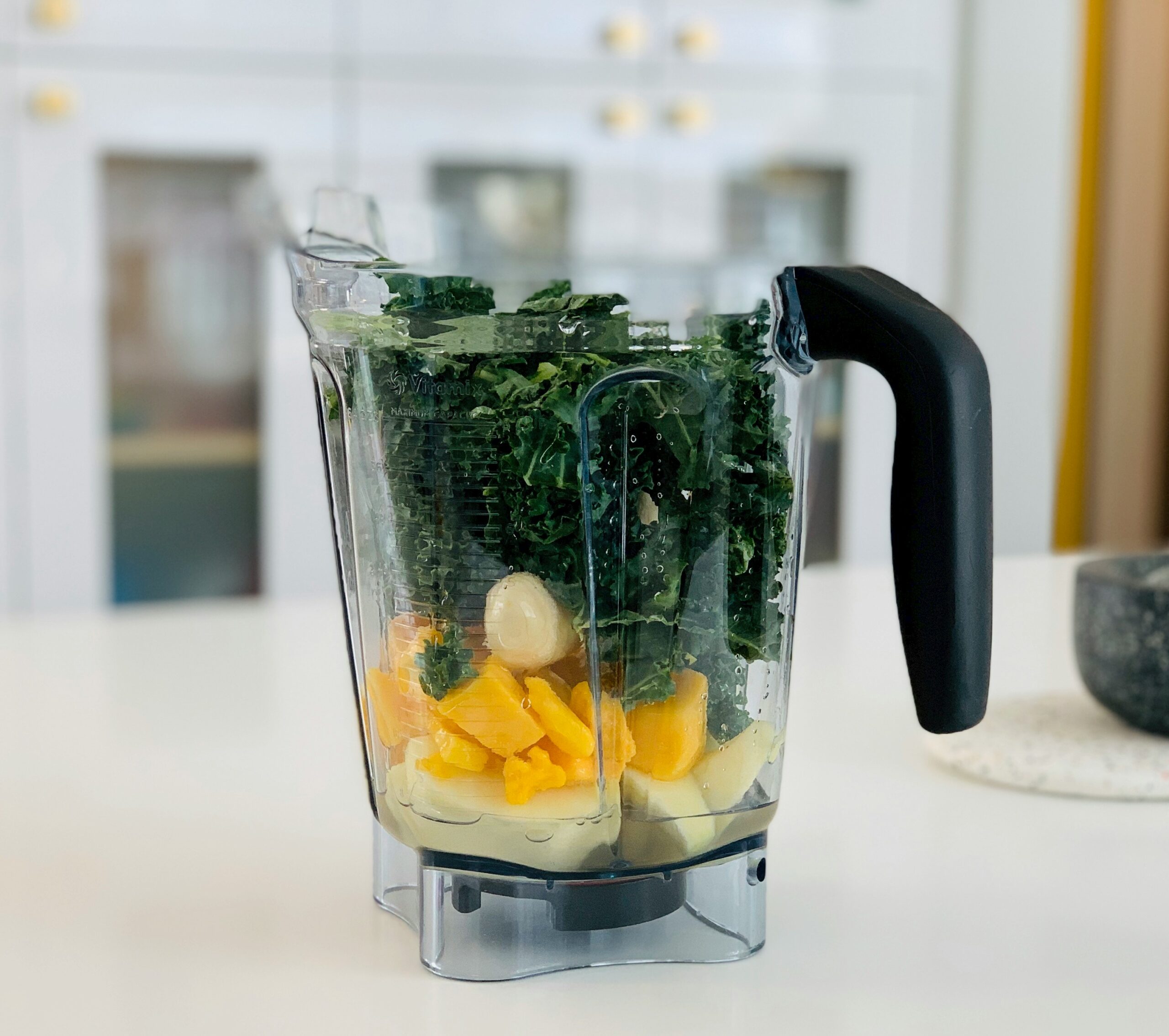
Blender Rumahan Vitamix A3500: Blender Premium Abdi Giri 22 April 2024 lewisart.biz – Bagi para pecinta healthy lifestyle, Vitamix adalah salah satu merek blender premium yang…
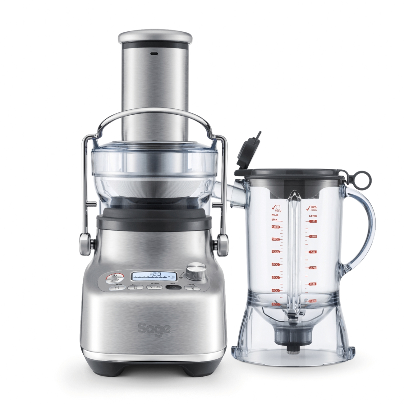
Blender Multifungsi Bluicer Blender & Juicer: Solusi Sempurna untuk Gaya Hidup Sehat Abdi Giri 4 April 2024 lewisart.biz – Dalam era modern di mana kesadaran akan gaya hidup sehat semakin meningkat, kebutuhan…
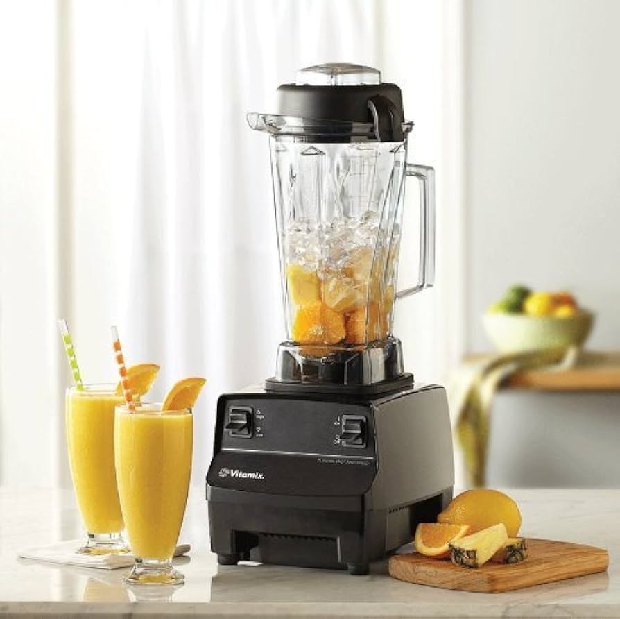
Blender Rumahan Blender Teknologi Canggih Kecepatan dan Kekuatan Blender TurboBlend 4500 Abdi Giri 26 March 2024 lewisart.biz – Dapur adalah pusat kreativitas di setiap rumah tangga, di mana makanan lezat dan…
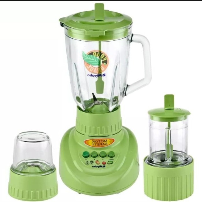
Blender Aman Mudah Digunakan Blender Rumahan Keunggulan Blender Miyako Abdi Giri 21 March 2024 lewisart.biz – Di era di mana kesehatan dan kebugaran semakin menjadi fokus utama, peralatan dapur…
Blender Rumahan Pilihan teratas untuk blender Cosmos Abdi Giri 13 March 2024 Anda mungkin familiar dengan Cosmos, sebuah merek yang menghasilkan berbagai perangkat elektronik berkualitas seperti rice…
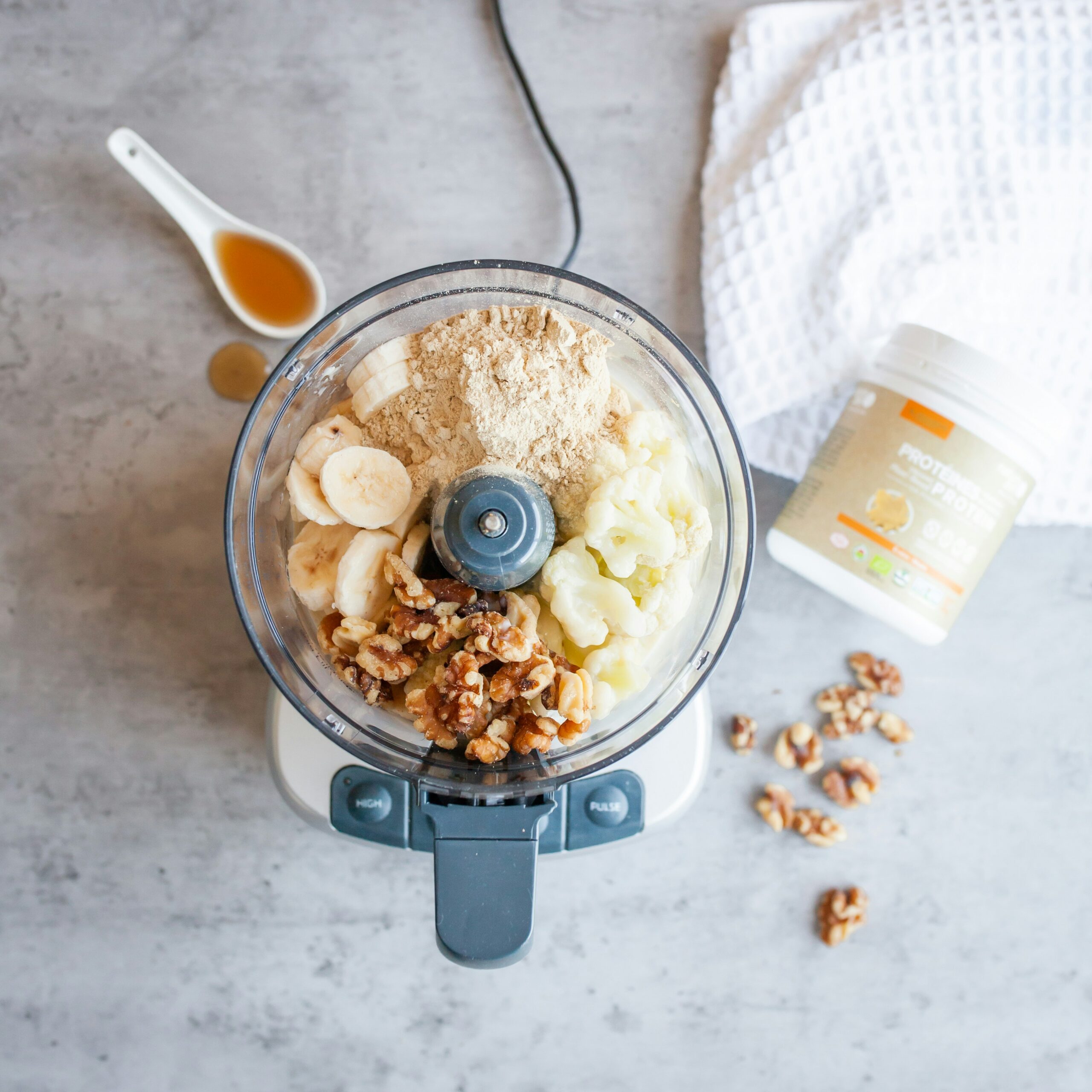
Blender Multifungsi Blender: Alat Multifungsi dalam Dapur Modern Abdi Giri 28 February 2024 lewisart.biz Blender merupakan salah satu peralatan dapur yang sangat penting dan multifungsi dalam rutinitas sehari-hari.…
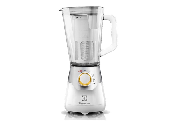
Blender Rumahan Electrolux Blender 2L EBR5604W, Usung 8 Multi Speed with Pulse Abdi Giri 4 September 2023 Electrolux Blender 2L EBR5604W bisa Anda gunakan untuk memenuhi berbagai keperluan. Mulai dari menggiling bumbu…
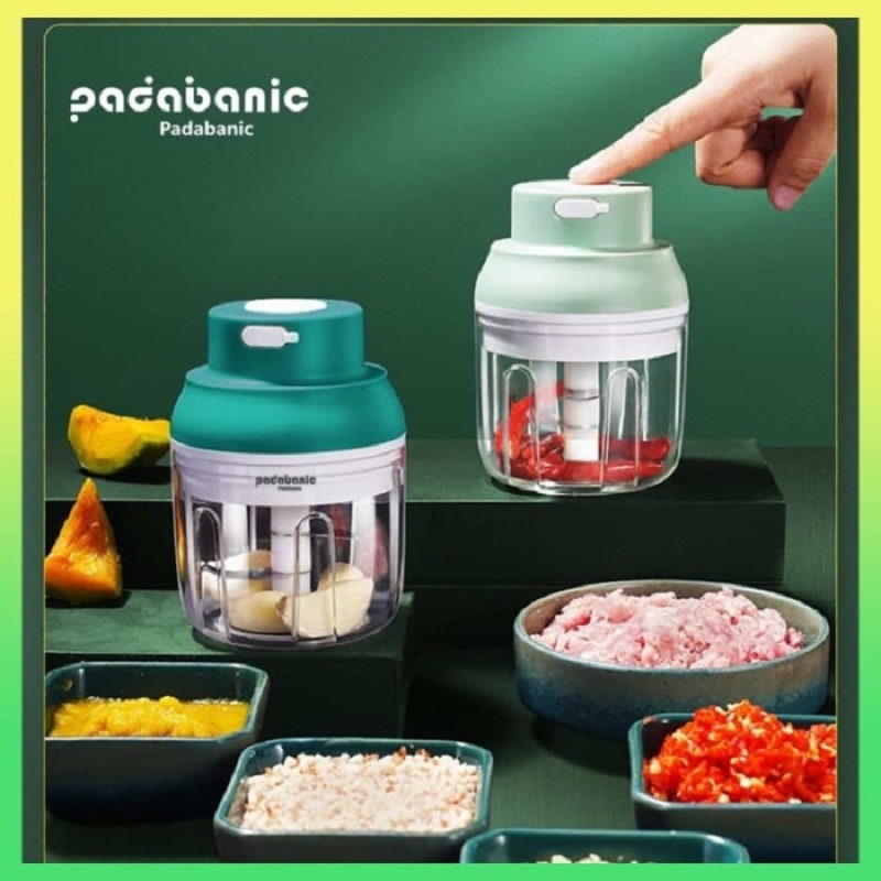
Blender Multifungsi Blender Penghalus Multifungsi PADABANIC, Praktis dan Terjangkau Abdi Giri 28 August 2023 Blender penghalus multifungsi PADABANIC perlu Anda miliki. Untuk Anda yang membutuhkan blender di rumah, produk…
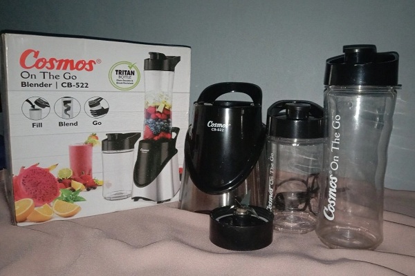
Blender Rumahan Cosmos CB 522 Blender Portable dengan Material Stainless Steel Abdi Giri 24 August 2023 Cosmos CB 522 dapat menjadi rekomendasi produk blender portable dengan berbagai rancangan fitur yang memudahkan…
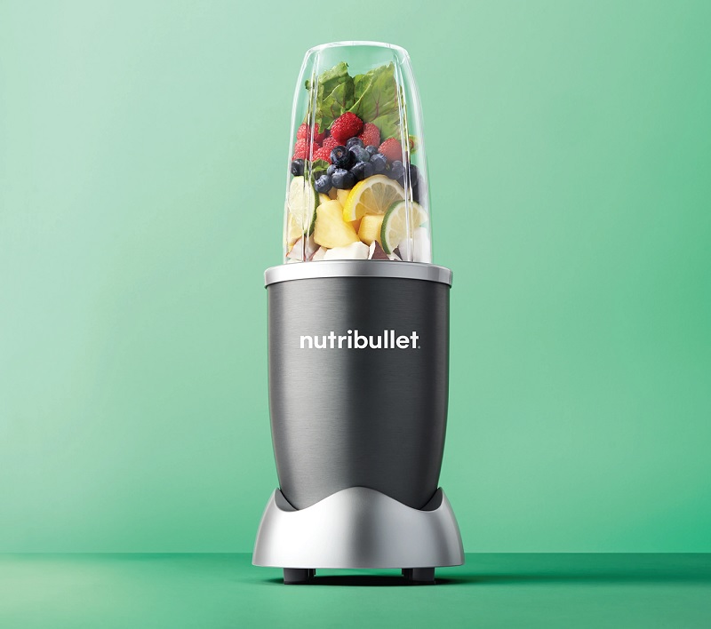
Blender Teknologi Canggih Blender Nutribullet 600 W Investasi Bagi Kesehatan yang Baik Abdi Giri 22 August 2023 Nutribullet 600 W blender nutrient extractor yang berasal dari Amerika Serikat. Dengan menggunakan blender tersebut,…